WEDNESDAY, April 30, 2014 (HealthDay News) — Doctors now can grow back large amounts of muscle lost to a traumatic injury, using tissue drawn from pigs as a “homing beacon” to coax the body’s own stem cells into repairing the wound.
Five patients with huge wounds in their leg muscles — including three injured during military service in Iraq and Afghanistan — experienced substantial regrowth following treatment with the pig tissue and intense physical therapy, a new study reported.
Three of the five patients had at least a 25 percent improvement in function following the treatment, and all five reported improved quality of life, the researchers noted.
The findings were published April 30 in Science Translational Medicine.
Trauma caused by a car crash or an explosive device can cause irreparable damage to a person’s muscles if too much muscle mass is torn away, said lead author Dr. Stephen Badylak, a surgical professor at the University of Pittsburgh and deputy director of the McGowan Institute for Regenerative Medicine.
“When you lose so much muscle that the gap is too large for the normal restorative processes to occur, the end result is typically filling that gap with scar tissue,” Badylak said in a Tuesday journal news briefing. The scar tissue causes a loss of function in that muscle, he said, potentially disabling the patient.
Badylak’s team hit upon the idea of using an “extracellular matrix” drawn from pig tissue to promote regrowth of muscle. Extracellular matrix is a component of body tissue that functions outside of the body’s cells. Made mostly of collagen, the extracellular matrix provides structural and biochemical support to surrounding cells.
Such materials already are used in hernia repair and breast reconstruction, to provide structural support and protection for surgical sites, study co-author Dr. Peter Rubin, chair of the department of plastic surgery at the University of Pittsburgh, said in the news briefing.
But researchers discovered that the implanted pig tissue promotes healing by releasing biochemicals called peptides into nearby human tissues.
“These peptides that are released serve as a homing device for the body’s own stem cells,” Badylak said. Stem cells located in nearby human tissues are drawn to the site of the wound, where they begin replacing the lost muscle.
After testing the process on mice, doctors started a clinical trial involving five men aged 27 to 37. All had lost between 58 percent to 90 percent of muscle in one of their legs.
The men underwent surgery that cut away all the scar tissue from the wound location, and then surgeons implanted the extracellular matrix into the wound.
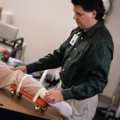
The patients all started aggressive rehabilitation within two days of the surgery, Badylak said. This was done to provide the stem cells guidance in the repair of muscle.
“When those cells get there, they depend upon local environmental cues to say, ‘OK, now that I’m here, what do you want me to do?’ One of the most important cues are the mechanical forces that are asked of the site,” he said.
The goal was to improve their ability to perform day-to-day tasks such as walking up stairs, getting out of a chair and raising a leg to a sitting position.
One of the patients, Nick Clark, lost massive amounts of muscle and nerve tissue from his left leg in a 2005 skiing accident. He had terrible balance in his left leg, he said, and sometimes relied on canes or ankle braces to stabilize his stride.
Clark underwent the experimental surgery in 2012. “The second day I was in the hospital, they had me walking up and down the halls of the hospital,” he said in the news briefing. “It was pretty tough. It was painful. But it was worth it.”
He now can balance on his left leg for several minutes at a time, and has more strength pushing off with his left foot.
“My balance is still not 100 percent, but it’s improved quite a bit,” said Clark, 34, of Youngwood, Penn. “Now I can almost keep pace with a normal person walking, maybe 90 percent. Before the surgery, I had about half the pace of a normal walk.”
The procedure represents an important medical advance, according to a trauma expert who was not involved in the study.
“I think it will set a new treatment paradigm in people with significant muscle injury, which we in the trauma world see quite frequently,” said Dr. David Lowenberg, an orthopedic surgeon at Stanford School of Medicine. “They took something that’s a huge problem and found a way to fix it relatively inexpensively, and it’s not technically difficult to do.”
Lowenberg helped train military doctors to deal with such large wounds as part of a distinguished scholars program, and saw firsthand the physical trauma caused by war. “The number of large muscle defects in our wounded warriors is real,” he said.
The cost of the procedure could decline even more in coming years. “There are new ways to manufacture these scaffolds that are even more cost-efficient and you won’t have to depend on the animals to process from,” Lowenberg said.
For now, study author Badylak said, the research serves as “a demonstration of the true movement of bench-top basic science through preclinical animal work to treatment at the bedside.”
He said the next step is to train surgical and physical therapy teams at other leading institutions on the process. “That way we can show this is an approach that can work,” Badylak said.
Once the procedure is proven effective, Badylak believes it could be used by any surgical hospital as a low-cost way to heal wounds that were previously thought irreparable.
“The approach we’ve taken is intended to be the type of approach that can be utilized anywhere that good surgery is available,” he said.
More information
The U.S. National Institutes of Health has more about stem cells.
Copyright © 2026 HealthDay. All rights reserved.

