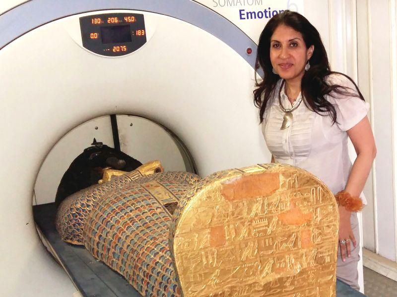
THURSDAY, Feb. 25, 2021 (HealthDay News) — Modern technology has unraveled an ancient mystery about the death of an Egyptian king.
Computed tomography (CT) scans of the mummified remains of Pharaoh Seqenenre Taa II, the Brave, revealed new details about his head injuries not previously found in examinations since his mummy was discovered in the 1880s. Those examinations, including an X-ray study in the 1960s, had found that the king had suffered several severe head injuries but no other wounds to his body.
Seqenenre briefly ruled over South Egypt during the country’s occupation by the Hyksos, a foreign dynasty that held power across the kingdom for about a century (c. 1650-1550 BC). He was killed during his attempt to oust the Hyksos. His violent death indirectly led to the reunification of Egypt in the 16th century BC.
Prior to the latest examination, the theory was that the king had been captured in battle and then executed afterward, possibly by the Hyksos king himself. It was also suggested he was murdered in his sleep by a palace conspiracy.
The new study now suggests that the king was captured on the battlefield, and then his hands were tied behind his back. The scans suggest the execution had been carried out by multiple attackers, according to the researchers. The scientists confirmed this by studying five different Hyksos weapons that matched the king’s wounds.
“This suggests that Seqenenre was really on the front line with his soldiers risking his life to liberate Egypt,” said lead author Dr. Sahar Saleem, a professor of radiology at Cairo University who specializes in paleoradiology.
“In a normal execution on a bound prisoner, it could be assumed that only one assailant strikes, possibly from different angles but not with different weapons,” Saleem explained. “Seqenenre’s death was rather a ceremonial execution.”
Detailed morphology revealed in the images made it possible for researchers to estimate that Seqenenre was about 40 when he died.
The findings were published Feb. 17 in the journal Frontiers in Medicine.
The study also revealed more information on the king’s mummification. While previously, the poor condition of the mummy suggested the embalming had been done hastily, away from the royal mummification workshop, the new research suggests it instead took place in a real mummification laboratory. The CT study revealed that the embalmers used a sophisticated method to hide the king’s head wounds under a layer of embalming material. This functioned similarly to the fillers used in modern plastic surgery.
Saleem and co-author Zahi Hawass, an archaeologist and former Egyptian minister of antiquities, have pioneered the use of CT scans to study the New Kingdom pharaohs and warriors. This includes well-known names such as Hatshepsut, Tutankhamun, Ramesses III, Thutmose III and Rameses II.
Saleem said the CT scan study provides important new details about a pivotal point in Egypt’s long history.
“Seqenenre’s death motivated his successors to continue the fight to unify Egypt and start the New Kingdom,” she said in a journal news release.
More information
The Smithsonian Institute has more information on Egyptian mummies.
SOURCE: Frontiers in Medicine, news release, Feb. 17, 2021
Copyright © 2026 HealthDay. All rights reserved.

